My favorite biology gifs
My favorite biology gifs
A study from the Dalia and Brun labs at Indiana University (go Hoosiers!) showed images of how Vibrio cholera can use pili to capture and take up nearby DNA. The videos of bacteria grabbing nearby DNA and the story of first author Courtney Ellison seeing the DNA uptake were picked up by the New York Times
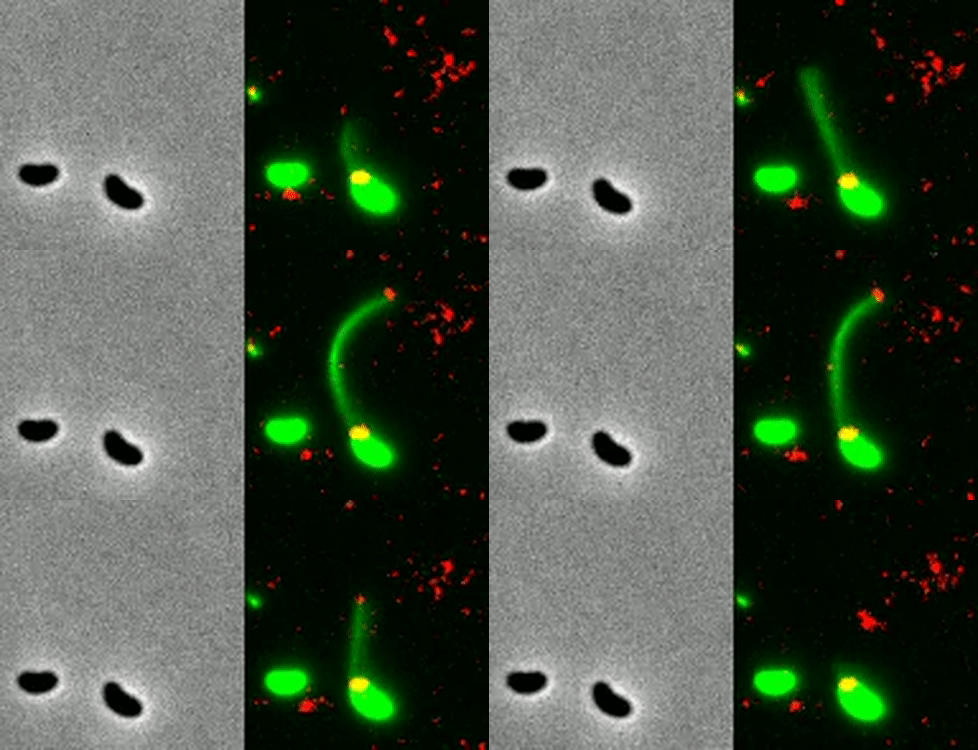
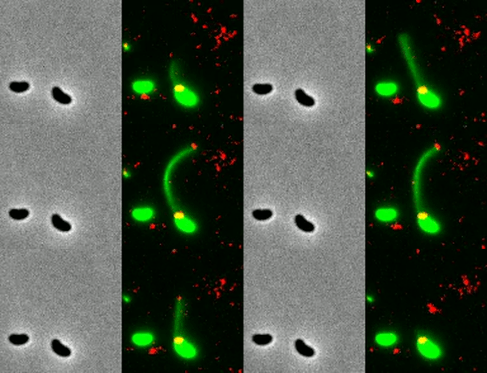
I first saw news of the work on Twitter from a university press release along with fantastic gif of a bacterium in action. A bacterial cell (labelled in green) uses a pilus to grab DNA (labelled red) and pulls it close to take it up. This was one of the most incredible videos of bacteria I’d ever seen!
So that got me thinking about some of my other favorite science gifs, so I decided to make a list. I made a few rules to narrow down from all of the science related gifs out there.
- Research video content only. There are lots of great animations or teaching content out there but here I’m focused on gifs created from lab research that let us witness science we hadn’t seen before. To this end, I’m including linked references for all of the gifs at the end.
- Biology only. This is mostly just due to what I know. I’d love to see the best space or physics or chemical reaction gifs but I’m certainly not the best person to make that list. In fact, I’m really focusing more on molecular and cellular biology here, so no wildlife gifs either or I would get carried away with large cats and capybaras.
- This is my list so at the end of the day it’s just my personal preference. Disagree? Make your own list! Or share it with me on Twitter @AaronJDy! I’d love to see new great science gifs.
1. Bacteria harpooning DNA
Here’s the recent gif that spurred me into compiling this list. It’s a really incredible look at a bacterium using a pilus to grab DNA several cell lengths away. Look that little guy grabbing that DNA! +1 for bacteria.
2. A white blood cell chasing bacteria
To me, this is the classic biology gif. In part because it’s created using film from the 1950s. In part because it shows the interplay of human cells and a bacterial pathogen. But mainly because it show cellular computation at such a direct level. A white blood cell chasing a bacterium through obstacles of red blood cells is no simple process and it paints a picture of an individual cell as much more complex than we typically give them credit for.
As a bioengineer I know that creating networks inside of a cell that can dynamically pursue and eliminate a moving target would not be easy so this little white blood cell also represents a goal to me for the kind of complexity that we will hopefully be able to one day engineer.


3. A human cell crawling in 3D space
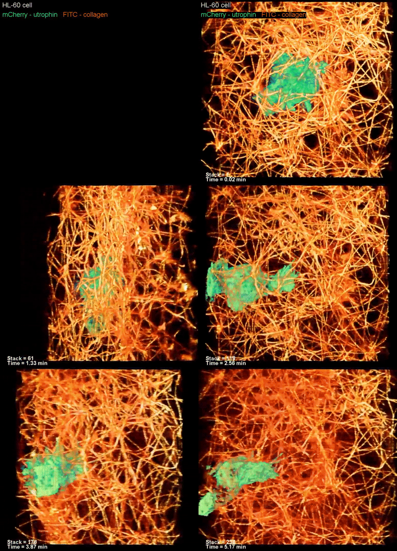
Here’s another look at how a human cell moves around. There’s no target to chase in this one but we get to see how it moves in all three spatial dimensions through a physical collagen network.

4. CRISPR-Cas9 cutting DNA
Now we’re getting pretty molecular. This gif is the often hyped CRISPR-Cas9 protein in action. This visualization comes from high speed atomic force microscopy. It’s a little grainy given the scale but see this oft talked about enzyme in action is pretty amazing.


5. Ribosome translocation
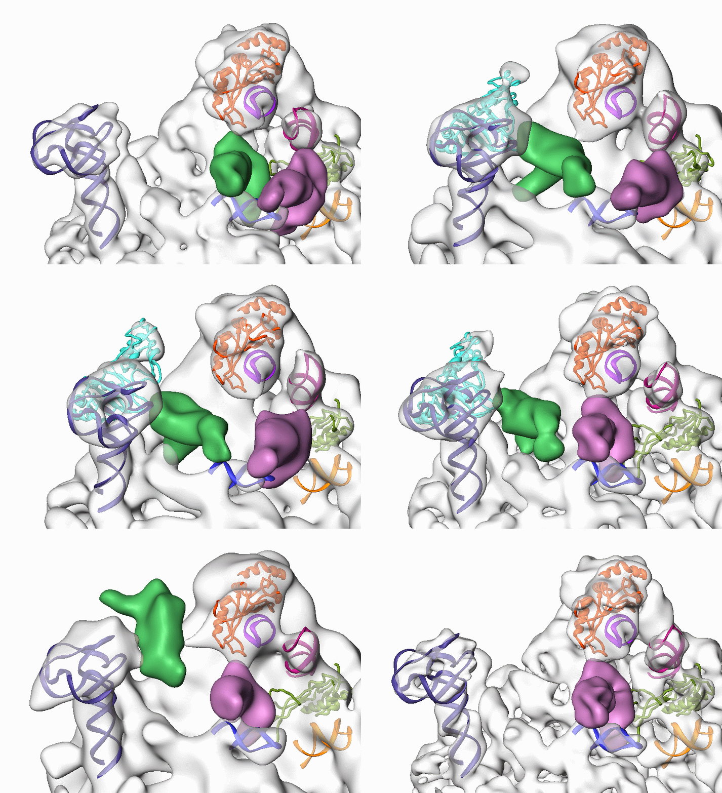
This gif is a visualization of a bacterial ribosome during translocation (movement of tRNAs along the three active sites of the ribosome) using electron cryomicroscopy. The grey structure is the 50S subunit and the 30S subunit has been digitally taken out so we can see the top of the 50S as the tRNAs are walked from site to site and making space for a new tRNA. There are many drawings and illustrations of translation but this really helps visualize what it looks like in 3D space.

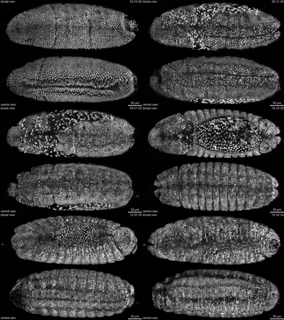
6. A developing fly embryo
Embryo development is another great category for being put in gif format. Such massive changes occur to cell patterning that it makes great videos when sped up. This makes use of light-sheet microscopy to image the entire embryo in real time. This technique uses a plane of light instead of a point so it can manage to image a full embryo much faster.

7. Zebrafish
Similar to the fly embryo, this is another use of light sheet micrscropy but looking at a zebrafish embryo. The paper behind this video imaged both wild-type and mutant zebrafish embryos over the first full day of development


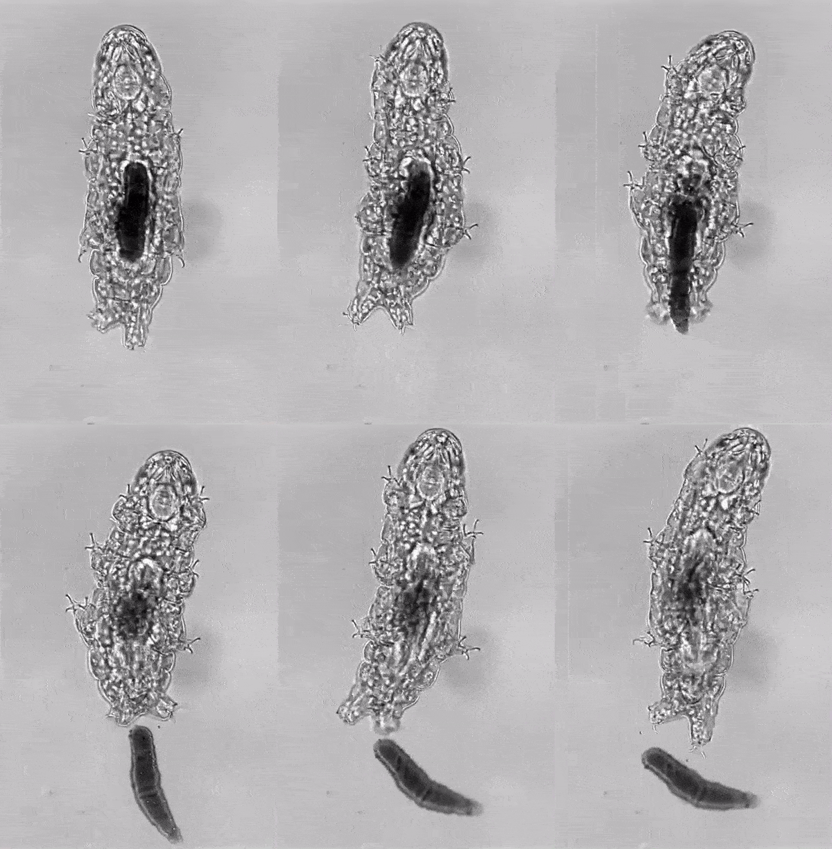
8. Tardigrade pooping
This one checks a lot of boxes. It involves the study tardigrade that have captured people’s imagination by being able to withstand harsh conditions of intense freezing, intense heat, complete dehydration, and radiation. It uses a microscope to catch details that the tardigrade seem like you should be able to see it with the naked eye. And maybe most importantly it involves a huge poo which people of all ages can find entertainment in.
I first saw this on Twitter from @TessaMontague after it went viral. You can read more here about how she captured the image at the MBL Embryology Course.

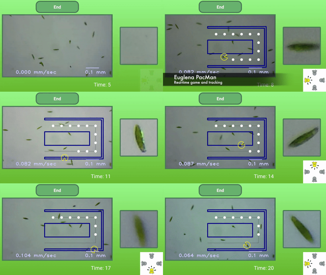
9. Biotic Pac Man game
I first saw the biotic games from Ingmar Riedel-Kruse’s lab when he came to give a talk at MIT. One of the tools to enable biotic games is called LudusScope which is a smartphone-based platform to interact with microscopic organisms. This gif is from a game using Euglena gracilis which are 50–80 micrometers in length and are phototactic (they move in response to light).
In this Pac Man game one cell is chosen to play. Then the game is overlaid on the screen with the live microscopy video. Four light controls are used to nudge the Pac Man cell in the direction you want it to go.
10. Horse gif encoded in DNA
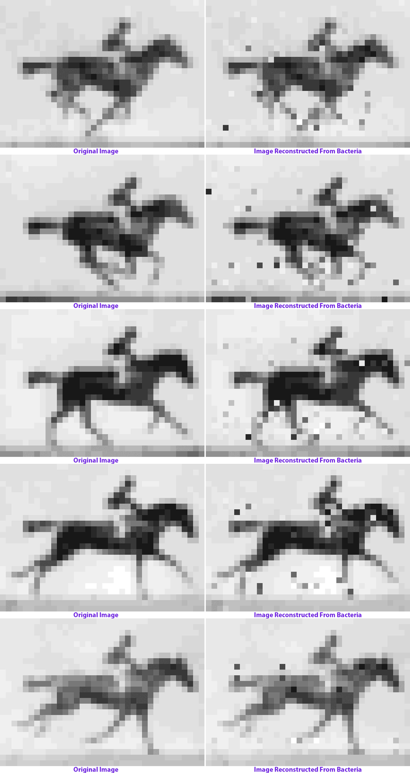
No fancy imaging here. Here a gif was the the goal. The pixel data for a gif was stored in DNA of living bacteria using CRISPR-Cas technology. The idea is to be able to store any kind of data in the genome but simple gifs were a fun example.
You can’t tell the bacteria is storing the information for a gif until you sequence the DNA and reconstruct the data you’ve stored.
I’m sure there are plenty of great biology gifs that I missed and even more science gifs in other disciplines. So let me know on Twitter @AaronJDy, share them with #sciencegifs, or make your own list of favorite science gifs!
